Liposomal Vitamin C Leukemia

Liposomal Vitamin C Leukemia

Photo Courtesy: Flashpop/DigitalVision//Getty Images

Whether in the form of a fizzy drink or flavored lozenges, cold and flu preventative supplements almost always highlight vitamin C as one of their key ingredients. So, what's so magical about vitamin C? Also known as ascorbic acid, vitamin C is critical to living healthily. Since the human body cannot spontaneously generate this nutrient, vitamin C must instead be absorbed from outside sources, such as vitamin supplements or foods that are naturally rich in it.

Commonly found in cold and flu preventative supplements, vitamin C strengthens and speeds up immune system functionality. Though research does not indicate that vitamin C intake alone can prevent the onset of cold or flu, adequate daily intake may shorten the duration of an infection or lessen the severity of symptoms.

Photo Courtesy: Catherine Falls Commercial/Moment/Getty Images

Vitamin C is crucial for the maintenance of well being. For example, it plays a role in wound healing and helps maintain many essential body tissues. It also acts as a potent antioxidant and can repair damage from free radicals, which are linked to aging effects, and disease vulnerability. Additionally, vitamin C can also prevent anemia, since it helps the body increase absorption of dietary iron, another vital mineral that the body cannot spontaneously create.

Foods that contain high concentrations of vitamin C have been linked with a lower risk of cardiovascular disease, like heart attack and stroke. Vitamin C can also increase levels of nitric oxide, a compound that widens blood vessels and, in turn, lowers blood pressure. In addition, regular intake of vitamin C, along with other vitamins, has been linked to a decreased risk for developing age-related cataracts, a leading cause of visual impairment in the United States.

Common Sources of Vitamin C

Vitamin C can be easily obtained through the many different foods, including:

Photo Courtesy: Akaradech Pramoonsin/Moment/Getty Images
  • Citrus fruits and juices (orange, grapefruit, lemon, lime and tangerine)
  • Berries
  • Melons
  • Mangoes
  • Kiwi
  • Tomato
  • Broccoli
  • Red peppers
  • Spinach
  • Squash
  • Potatoes

Cooking these foods may result in the loss of some of the vitamin content, so it is ideal to ingest them raw, either whole or juiced. Nowadays, there are also numerous packaged food products, like cereals, that have been enriched and fortified with vitamin C, so that the nutrient can be easily obtained.

Vitamin C may also be labeled as "L-ascorbic acid" in supplement form, and most over-the-counter multivitamins contain the recommended daily amount of the vitamin. While it is a good source when an individual is in need of a vitamin C boost, supplements are not meant to replace a diet rich in naturally derived vitamin C.

What Happens When You Have Too Much — or Too Little — Vitamin C?

Vitamin C is a water-soluble vitamin that can be easily flushed out of the body via urination when it is not needed. Therefore, if the main source of vitamin C is from naturally occurring foods, it is near-impossible for excess vitamin C to produce side effects. However, taking excessive concentrated vitamin C supplements may lead to diarrhea or stomach upset.

Photo Courtesy: Violeta Stoimenova/E+/Getty Images

Since vitamin C-rich foods are so readily available nowadays, symptoms of inadequate vitamin C intake are also rare in the United States. However, malnourished individuals can experience symptoms of vitamin C deficiency over time, including:

  • Weakness
  • Fatigue
  • Anemia
  • Easy bruising
  • Joint pain
  • Skin breakdown
  • Weakened tooth enamel
  • Gum inflammation

Severe vitamin C deficiency is referred to as scurvy. Scurvy can be easily treated with increased dietary or supplemental vitamin C. Since vitamin C is crucial in the detoxification of the body, a lack of vitamin C can compromise the immune system and make an individual more susceptible to diseases and infections. Individuals with insufficient vitamin C may find that it takes longer than usual to recover from a cold or a physical wound.

Daily Dosage Recommendations:

The daily dosage recommendation for vitamin C is different for everyone, depending on factors such as gender, age, lifestyle and current health condition. The recommended daily dosage for vitamin C is at least 75 mg daily for women and 90 mg for men. Since people who are pregnant, breast feeding, smoking or using oral contraceptives have a lower blood level of vitamin C than others, larger doses of vitamin C may be needed to achieve optimal results in these individuals. Those who have prior or current medical conditions may also require bigger or smaller dosage levels, as recommended by their healthcare providers.

Resource Links:

  • "Vitamin C" via MedlinePlus
  • "Vitamin C and Infections" via MDPI
  • "Extra Dose of Vitamin C Based on a Daily Supplementation Shortens the Common Cold: A Meta-Analysis of 9 Randomized Controlled Trials" via Hindawi, BioMed Research International
  • "Vitamin C" via National Institutes of Health
  • "Scurvy" via U.S. Department of Health & Human Services, National Institutes of Health
  • "Dietary intake and blood concentrations of antioxidants and the risk of cardiovascular disease, total cancer, and all-cause mortality: a systematic review and dose-response meta-analysis of prospective studies" via The American Journal of Clinical Nutrition
  • "Dietary vitamin and carotenoid intake and risk of age-related cataract" via The American Journal of Clinical Nutrition
  • "Cardiovascular System" via Department of Anatomy, Seoul National University College of Medicine (via Springer)

MORE FROM SYMPTOMFIND.COM

Liposomal Vitamin C Leukemia

Source: https://www.symptomfind.com/health/vitamin-c-everything-you-need-to-know?utm_content=params%3Ao%3D740013%26ad%3DdirN%26qo%3DserpIndex

Share:

How Much Vitamin C Is In Cantaloupe

How Much Vitamin C Is In Cantaloupe

Cantaloupes: Health Benefits & Nutrition Facts

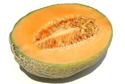
(Image credit: stock.xchng)

A sweet, juicy slice of cantaloupe is refreshing on a hot summer day, or any time of year. Like other melons, cantaloupe has a high water content (about 90 percent), but being packed with water doesn't mean that this popular fruit lacks nutritional value.

In fact, a cantaloupe is bursting with nutrients: It's loaded with vitamin A (in the form of beta-carotene) as well as vitamin C, and is a good source of the mineral potassium. Another benefit is that the fruit's deep-orange flesh is full of flavor, but is low in calories.

"This melon is a great choice when it comes to nutrients per calorie," said Heather Mangieri, a Pittsburgh-based registered dietitian and nutritionist, author and spokesperson for the Academy of Nutrition and Dietetics.

"One cup of cantaloupe contains only about 55 calories (due to its high water content) but offers over 100 percent of your daily needs for vitamin A, over 50 percent of the daily needs for vitamin C, 1.5 grams of fiber and is a good source of potassium," Mangieri said.

What's more, a 2006 study published in HortScience found that cantaloupes have even higher concentrations of beta-carotene, which are plant pigments found in yellow and orange fruits and vegetables that the body converts to vitamin A, than oranges, even though oranges are brighter in color. Cantaloupe is one of the best sources of vitamin A among fruits and the top source among melons.

Nutrition facts

Here are the nutrition facts for one cup of cantaloupe, according to the U.S. Food and Drug Administration:

Cantaloupe Serving size: 1 cup, cubed (160 g) Calories 54 (Calories from Fat 3) *Percent Daily Values (%DV) are based on a 2,000 calorie diet.

Nutrition Facts Cantaloupe Serving size: 1 cup, cubed (160 g) Calories 54 Calories from Fat 3 *Percent Daily Values (%DV) are based on a 2,000 calorie diet. Amt per Serving %DV* Amt per Serving %DV*
Total Fat 0g 0% Total Carbohydrate 14g 5%
Cholesterol 0mg 0% Dietary Fiber 1g 6%
Sodium 26mg 1% Sugars 13g
Protein 1g
Vitamin A 108% Calcium 1%
Vitamin C 98% Iron 2%

About melons

It is thought that the fruit was named "cantaloupe" for Cantalupo, an Italian town near Vatican City, where melon seeds brought from Armenia were planted in the papal gardens during the Renaissance, according to World's Healthiest Foods.

Cantaloupes are in the Cucurbitaceae, or gourd family, which includes other plants that grow on a vine, such as watermelon, honeydew and casaba melons, as well as pumpkins, squash and cucumbers.

According to the University of Illinois Extension, the cantaloupe is a variety of muskmelon. North American cantaloupes (Cucumis melo reticulatus) are known for their uniform "netting" over the rind; European cantaloupes (Cucumis melo cantalupensis) have greener skin, little netting, deep grooves and would surprise most Americans by being called cantaloupes.

Picking a ripe one

Selecting a fresh cantaloupe can be tricky because you can't see the inside the melon.  But according to Mangieri, freshness is critical to the fruit's sweet flavor. Pick up a cantaloupe and if it feels heavier than you expected, it's likely ripe. A ripe melon should smell sweet when you place your nose next to the fruit, and you should be able to push in the skin a little bit with your thumb.

If the melon is not quite ripe when you buy it, you can set it on a kitchen counter for a few days. But don't wash the fruit at this point — wait until you're ready to cut the melon to wash its outer surface to reduce the chance for bacterial growth.

"While a cantaloupe will become softer and juicer with time, the fruit's sugar content [and sweetness] will not significantly increase after it is harvested," Mangieri told Live Science.

Health benefits

Cantaloupe is not a well-studied fruit on its own. Most of the research on the health benefits of the melon has focused on a person's total dietary intake of fruits and vegetables in general, or studies have looked at diets rich in specific nutrients or plant compounds found in these fruits, such as carotenoids, potassium or vitamin C. This makes it hard to draw firm conclusions about the unique health benefits of cantaloupe.

Antioxidant power

Cantaloupe is a rich food source of vitamins A and C.

"Vitamins A and C are both antioxidants that work to keep your body healthy," Mangieri said. Antioxidants can have protective effects by neutralizing free radicals, which can damage DNA in cells and promote chronic inflammation in the body.

Free radicals cause cell damage and disruption that can contribute to diseases. "[Antioxidants such as vitamins A and C] may help prevent conditions such as cancer, heart disease, and arthritis," Mangieri added.

Heart health

There is strong evidence that a diet rich in fruits and vegetables is linked with a reduced risk of heart disease and stroke, and can also lower blood pressure, according to the Harvard T.H. Chan School of Public Health.

Eye health

Including more fruits and vegetables in your diet can keep your eyes healthy and may help fend off cataracts and macular degeneration, two common age-related eye problems, according to the Harvard T.H. Chan School of Public Health.

The vitamin A found in cantaloupe is a key nutrient for good vision, Mangieri said.

Digestion

The fiber and water in cantaloupe can aid digestion and help prevent constipation, when included as part of a high-fiber diet, such as a diet rich in fruits, vegetables and whole grains, according to the Mayo Clinic.

Risks of eating cantaloupe

In general, enjoying cantaloupe poses little risk for most people. However, cantaloupes have been linked to more than 10 foodborne illness outbreaks in the past 10 to 15 years, according to the Centers for Disease Control and Prevention (CDC). The majority of these incidents were bacterial infections caused by salmonella, but people have also been sickened by E.coli, and there were some deaths reported in a multistate outbreak of listeria.

In one analysis published in Epidemiology and Infection in 2006, researchers found that 25 outbreaks were linked to the consumption of cantaloupe and reported to the CDC between 1973 and 2003. These outbreaks affected more than 1,600 people, but the researchers suspect that the actual number of people sickened by eating contaminated cantaloupe was probably much higher because some cases of cantaloupe-related illness may never have been reported to health officials.

Cantaloupe may be vulnerable to outbreaks of foodborne illness because the fruit is grown in close contact with the ground, where it may become contaminated with bacteria from the soil, water or animals before it is harvested, according to Colorado State University. In addition, the melons have a rough and textured outer surface that can trap bacteria. Bacteria can also be transmitted during the processing of pre-cut melon, from a knife cutting through contaminated rinds. If the same contaminated knife continues to be used, it can transfer bacteria to the flesh inside. (To stay safe when cutting cantaloupe at home, see the tips below.)

Bacterial contamination is not the only possible risk from eating cantaloupe. Some people with allergies to ragweed pollen may also develop symptoms of oral allergy syndrome immediately after eating melons, such as cantaloupe, watermelon or honeydew.

When some people who experience ragweed allergies start to eat cantaloupe, they may get an itchy feeling in their throats and lips or have swelling in their mouths, tongues and throats. This reaction occurs because the body's immune system recognizes a similarity between the allergy-causing proteins in ragweed pollen and the proteins in the food. (Besides melons, ragweed sufferers may also be sensitive to kiwi, banana, cucumber and zucchini.)

Tips for cutting cantaloupe

  • Purchase melons without any visible bruises, cracks or soft spots on the skin.
  • Wash hands with soap and water before handling cantaloupes.
  • Scrub the outer surface of the melon with a vegetable brush under cool tap water before eating the fruit. Pat the fruit dry with paper towels to remove excess water.
  • Using a clean knife and cutting board, cut off the stem end (where the fruit was attached to the vine) of the cantaloupe and throw it out. Studies have found that this area is most likely to have bacterial contamination.
  • Cut the entire melon in half and scoop out the seeds and strings. Using a knife or melon baller, cut up the orange flesh.
  • After cutting up the melon, wash any utensils and cutting boards used in hot, soapy water and refrigerate the sliced melon.

Fun facts

Cantaloupe seeds can be roasted and eaten as a snack.

California is the largest cantaloupe-producing state in the country. Over half of all U.S. cantaloupes are grown there. The next six states are Arizona, Colorado, Georgia, Indiana and Texas.

The United States also imports cantaloupes each year, primarily from Guatemala, Honduras, Costa Rica and Mexico.

Additional reporting by Cari Nierenberg, Live Science contributor.

Additional resources

  • University of Illinois Extension: Cantaloupe or Muskmelon?
  • World's Healthiest Foods: Cantaloupe
  • University of Maine Extension: Melons

How Much Vitamin C Is In Cantaloupe

Source: https://www.livescience.com/54475-cantaloupe-nutrition.html

Share:

Highest Vitamin C Foods In Order

Highest Vitamin C Foods In Order

vitaminc-mango

Credit: Getty Images

Load your plate with these vitamin C-rich eats, which some research shows can build your body's defense against colds and the flu.

1 of 13

Foods high in vitamin C

vitaminc-orange

Credit: Getty Images

Advertisement

Advertisement

2 of 13

Chili peppers

vitaminc-chili-peppers

Credit: Getty Images

A half-cup of chopped or diced chili peppers delivers 107.8 mg of vitamin C. Plus, researchers from the University of Buffalo found that capsaicin, the compound that makes chili peppers hot, may help relieve joint and muscle pain.

RELATED: Is It Dangerous to Eat Really Hot Peppers?

3 of 13

Red bell pepper

vitaminc-peppers

Credit: Getty Images

Advertisement

4 of 13

Green bell pepper

vitaminc-greenpepper

Credit: Getty Images

A cup of chopped green bell pepper contains less vitamin C than its sweeter sister, but at 120 mg, it's still 200% of your recommended daily allowance. Green bell pepper is also a great source of fiber.

RELATED: 10 Keto-Friendly Vegetables You Should Eat More Of

5 of 13

Kale

vitaminc-kale

Credit: Getty Images

6 of 13

Broccoli

vitaminc-broccoli

Credit: Getty Images

Advertisement

Advertisement

7 of 13

Papaya

vitaminc-papaya

Credit: Getty Images

8 of 13

Strawberries

vitaminc-strawberries

Credit: Getty Images

9 of 13

Cauliflower

vitaminc-cauliflower

Credit: Getty Images

Advertisement

10 of 13

Brussels sprouts

vitaminc-sprouts

Credit: Getty Images

These little cabbages are loaded with cancer-preventing phytonutrients and fiber, not to mention 74.8 mg of vitamin C. If you're usually turned off by their bitter taste, bring out their natural sweetness by roasting them.

RELATED: 7 Health Benefits of Brussels Sprouts

11 of 13

Pineapple

vitaminc-pineapple

Credit: Getty Images

In addition to 78.9 mg of vitamin C, pineapple contains bromelain, a digestive enzyme that helps break down food and reduce bloating. Bromelain also acts as a natural anti-inflammatory that can help you recover faster after a tough workout.

RELATED: The Best and Worst Foods for Your Vagina

12 of 13

Kiwi

vitaminc-kiwi

Credit: Getty Images

Advertisement

13 of 13

Mango

vitaminc-mango

Credit: Getty Images

Taste the tropics for a 122.3 mg boost of vitamin C. Mango is also a great source of vitamin A, which like vitamin C plays a key role in immunity and additionally keeps your eyes healthy.

RELATED: 6 Health Benefits of Mangoes

To get our top stories delivered to your inbox, sign up for the Balanced Bites newsletter

Up Next

Highest Vitamin C Foods In Order

Source: https://www.health.com/nutrition/12-foods-with-more-vitamin-c-than-oranges

Share:

Garden Of Wisdom Vitamin C Ferulic

Garden Of Wisdom Vitamin C Ferulic

Researchers in the United Kingdom say a study on eyesight shows diet and environmental factors are more important than genetics in lowering risk of cataracts.

Eating an apple a day may keep the doctor away, but eating oranges might do the same for cataracts.

In a study published today in the journal Ophthalmology, researchers in the United Kingdom said a higher dietary intake of vitamin C might significantly reduce the risk of developing cataracts.

The study, led by scientists at King's College London, is the first to conclude that dietary and environmental factors play a larger role than genetics in the development of cataracts.

"The findings of this study could have significant impact, particularly for the aging population globally by suggesting that simple dietary changes such as increased intake of fruits and vegetables as part of a healthier diet could help protect them from cataracts," Dr. Chris Hammond, professor of ophthalmology at King's College, consultant eye surgeon and lead author of the study, said in a statement.

Read More: What Is a Cataract? »

The researchers estimated genetic factors account for 35 percent of the difference in cataract progression. Environment and lifestyle account for 65 percent.

To study the impact diet has on cataracts, the researchers tracked the progression of the eye condition in 324 pairs of female twins from the United Kingdom.

The scientists examined digital images of the women's eye lenses when they were about 60 years old. They then studied the same type of images 10 years later.

They kept track of the women's intake of vitamins A, B, C, D, and E. They also tracked their intake of copper, manganese, and zinc using a food questionnaire.

The researchers said the women who ingested more vitamin C initially had a 20 percent reduced risk of developing cataracts. After 10 years, that risk had decreased by 33 percent.

The researchers noted that there was little risk reduction in the women who took vitamin supplements. Instead the preventative effects appeared to be obtained only by eating foods rich in vitamin C.

Dr. Ravi D. Goel, an ophthalmologist from New Jersey who is also a clinical instructor at Wills Eye Hospital in Pennsylvania, said the study provides helpful information for patients and doctors.

"These are novel findings for patients going forward," Goel, a spokesperson for the American Academy of Ophthalmology, told Healthline. "This is a helpful tool for patient education."

Read More: Americans Spend Billions on Vitamins and Supplements That Don't Work »

Cataracts occur when the lens of the eye becomes cloudy due to oxidation over a long period of time.

The researchers said the fluids that bathe the eye are rich in vitamin C, which helps stop the lens from oxidizing.

The dietary intake of vitamin C helps prevent cataracts by increasing the amount of this vitamin in the eye fluid.

The researchers added that smoking and diabetes also are risk factors for certain kinds of cataracts, so a balanced diet and healthy lifestyle are important.

"Healthy diets are always an advantage for patients," added Goel.

Goel also said vitamin C has already been shown to help slow the progression of age-related macular degeneration.

This latest information on cataracts adds to vitamin C's attributes. "It helps overall eye health," he said.

The researchers did note that their observational study has its limitations as it only involved women who were aged 60 years and older.

However, the researchers believe the information could also be relevant for male patients.

Cataracts are the leading cause of blindness in the world, affecting about 20 million people, according to statistics from the World Health Organization (WHO). Cataracts also affect 24 million Americans over the age of 40.

The condition can cause blurry vision, glare, poor night vision, and sensitivity to light.

Initially, better lighting and glasses may help ease some of the symptoms, but as cataracts progress surgery is sometimes needed.

Read More: Diabetes and Blurry Vision: What You Need to Know »

Garden Of Wisdom Vitamin C Ferulic

Source: https://www.healthline.com/health-news/vitamin-c-may-reduce-risk-of-cataracts

Share:

Emergen C Vitamin Powder

Emergen C Vitamin Powder

Photo Courtesy: KARRASTOCK/Moment/Getty Images

Nearly 2 million American adults each year are diagnosed with cancer. If you receive a diagnosis, your schedule can quickly start to fill up with doctor's appointments, medical procedures and pharmacy visits. Creating a checklist of things to do can help keep you busy and feeling more in control. But acknowledging the effects a cancer diagnosis can have on your emotions — and healthily managing those emotions — is almost as important.

Doctors and psychologists now recognize that healing improves after a diagnosis when both our physical and emotional needs are met, says Niki Barr, Ph.D., a psychotherapist at the Center for Cancer and Blood Disorders in Texas, who consults with medical doctors, extended families and caregivers about emotionally coping with a cancer diagnosis.

Learning you have cancer is a stressful experience, and it's important to acknowledge and cope with that stress. Fully understanding how your emotional needs can change after you receive a cancer diagnosis can go a long way in helping you and your family heal. The tips here can help you support your mental health as you navigate your diagnosis and treatment.

Photo Courtesy: burakkarademir/E+/Getty Images

Upon hearing your diagnosis, you might experience grief, fear or even a feeling of denial until some time has passed. "These are all normal and emotionally healthy responses, but it's all too easy to spiral out of control with fear in the beginning," says Barr. Fortunately, these tips can help you manage the anxiety and fear that come along with learning about a cancer diagnosis.

Lessen the Impact of Anxious Thoughts

Start by writing your thoughts down on note cards or in a journal. Identify the first one that's leading you to feel uneasy. For example, you might write down something like "I'm afraid of my hair falling out." Then, move onto the subsequent fear and write it down. That might be something like "People will treat me differently if they see I have cancer."

When you've identified most of your anxious thoughts, go back to the first one and write something new on the card that can help ease your stress. It should be a thought that's confident and empowering. For example, suppose you're worried about your hair falling out. In that case, a positive view could be: "I've been looking forward to getting a new hairstyle anyway." When you're feeling nervous, read the more positive strategies, says Barr.

Work On Your Internal Dialogue

It can also help to defuse all those "What if?" questions you might ask yourself, such as "What if my cancer has spread?" or "What if the treatment doesn't work?" One scary question tends to lead to another and often turns into full-fledged anxiety. Try to focus on those things you have the power to improve on your own. The next time you start asking yourself the what-ifs, substitute the upsetting ideas with this one: "Is this thought helping me or hurting me?" You can also ask, "Is this thought moving me forward or backward?" Your answers might surprise you, and they can help you move away from the thoughts that are holding you back.

Get Grounded

Interrupting periods of anxiety by focusing on small details around you can help you shift your perspective away from negativity and towards the present moment. "Look at the beautiful color of the walls in the room you're in; look at the person you're talking to, the clothes [they're] wearing," Barr suggests. Becoming very present and focused on physical details nearby helps soothe sudden anxious thoughts. Turn your focus towards absorbing the colors, smells, people and each new sound around you. Build those sensations up very clearly in your mind. You can use this technique as a distraction tool the next time you're waiting for a medical procedure or want a diversion from your thoughts.

Meditate to Music

Research shows that 15 to 30 minutes of both guided imagery and soothing tunes can alleviate deep feelings of stress about a cancer diagnosis. The Cancer Treatment Centers of America (CTCA) reviewed 30 clinical cancer trials that analyzed more than 1,890 cancer patients. They found that music therapy can have a beneficial effect on anxiety, pain, mood, quality of life, heart rate, respiratory rate and blood pressure.

The CTCA also offers many classes and therapies to help people relax, reduce stress and improve their quality of life as part of an extensive mind-body medicine program. Many centers provide calming background music during healing therapies.

What Are Your Next Steps?

Photo Courtesy: Peathegee Inc/Getty Images

You have a cancer diagnosis: Now what? Learning how to clear your mind and focus on positive thoughts is a helpful step in the right direction. Here are other soul-soothing strategies you can try.

Start Journaling and Reflecting

Research published in the Journal of Clinical Oncology has shown that expressing your innermost feelings can reduce stress and have a range of other emotional and social benefits. Researchers aren't sure why putting thoughts down on paper is effective. Still, it allows you to process complex emotions and help you chart a way forward, whether you've been diagnosed with cancer or are taking care of someone who has.

Exercise When Possible

Exercising for 2.5 hours per week can help you beat symptoms of depression and fatigue. Among the nation's millions of cancer survivors, there are hints — but not proof yet — that active exercisers may lower their risk of their cancer coming back.

The American College of Sports Medicine hosted a medical panel of cancer specialists to evaluate this exercise evidence. It issued guidelines suggesting that cancer patients and survivors should exercise for about 30 minutes most days of the week. This exercise should be enjoyable to you but also build up a sweat. When patients and their family caregivers exercised together, research found, everyone was more likely to stick with the fitness regimen, boost their physical stamina and experience less emotional strain.

Strengthen Your Social Support System

Connecting with others who've been through this kind of emotional diagnosis already can be a source of comfort and support. Learn more about online communities and your local chapters and support groups that meet up. The American Cancer Society also suggests attending one-on-one professional therapy or its "I Can Cope" online support groups to learn more.

Resource Links:

Find Local Cancer Support Programs | Cancer Support Groups

Strategies Used in Coping With a Cancer Diagnosis Predict Meaning in Life for Survivors

Coping Well with Advanced Cancer: A Serial Qualitative Interview Study with Patients and Family Carers

Coping with cancer

Coping Attitudes of Cancer Patients and Their Caregivers and Quality of Life of Caregivers

MORE FROM SYMPTOMFIND.COM

Emergen C Vitamin Powder

Source: https://www.symptomfind.com/health/tips-coping-with-cancer-diagnosis?utm_content=params%3Ao%3D740013%26ad%3DdirN%26qo%3DserpIndex

Share:

Does Vitamin C Increase Breast Size

Does Vitamin C Increase Breast Size

Vitamin C promotes apoptosis in breast cancer cells by increasing TRAIL expression

David W. Sant

1John P. Hussman Institute for Human Genomics, Dr. John T. Macdonald Foundation Department of Human Genetics, University of Miami Miller School of Medicine, Miami, FL 33136 USA

Sushmita Mustafi

1John P. Hussman Institute for Human Genomics, Dr. John T. Macdonald Foundation Department of Human Genetics, University of Miami Miller School of Medicine, Miami, FL 33136 USA

Christopher B. Gustafson

1John P. Hussman Institute for Human Genomics, Dr. John T. Macdonald Foundation Department of Human Genetics, University of Miami Miller School of Medicine, Miami, FL 33136 USA

Joshua Chen

1John P. Hussman Institute for Human Genomics, Dr. John T. Macdonald Foundation Department of Human Genetics, University of Miami Miller School of Medicine, Miami, FL 33136 USA

Joyce M. Slingerland

2Braman Family Breast Cancer Institute, University of Miami Miller School of Medicine, Miami, FL 33136 USA

3Sylvester Comprehensive Cancer Center, University of Miami Miller School of Medicine, Miami, FL 33136 USA

Gaofeng Wang

1John P. Hussman Institute for Human Genomics, Dr. John T. Macdonald Foundation Department of Human Genetics, University of Miami Miller School of Medicine, Miami, FL 33136 USA

3Sylvester Comprehensive Cancer Center, University of Miami Miller School of Medicine, Miami, FL 33136 USA

Received 2018 Jan 5; Accepted 2018 Mar 19.

Abstract

Genomic loss of 5-hydroxymethylcytosine (5hmC) accompanies malignant cellular transformation in breast cancer. Vitamin C serves as a cofactor for TET methylcytosine dioxygenases to increase 5hmC generation. Here we show that the transcription of SVCT2, a major vitamin C transporter, was decreased in human breast cancers (113 cases) compared to normal breast tissues from the same patients. A decreased SVCT2 expression was also observed in breast cancer cell lines. Treatment with vitamin C (100 μM) increased the 5hmC content in MDA-MB-231 breast cancer cells and markedly altered the transcriptome. The vitamin C treatment induced apoptosis in MDA-MB-231 cells, which was verified in two additional breast cancer cell lines. This pro-apoptotic effect of vitamin C appeared to be mediated by TRAIL, a known apoptosis inducer. Vitamin C upregulated TRAIL transcripts (2.3-fold increase) and increased TRAIL protein levels. The upregulation of TRAIL by vitamin C was largely abolished by siRNAs targeting TETs and anti-TRAIL antibody abrogated the induction of apoptosis. Furthermore, the apoptosis promoted by vitamin C was associated with Bax and caspases activation, Bcl-xL sequestration, and cytochrome c release. Taken together, these results suggest a potential role of physiological doses of vitamin C in breast cancer prevention and treatment.

Introduction

Aberrant epigenetic alterations, which reflect the interface of a dynamic microenvironment and the genome are involved in malignant cellular transformation 1 . Global loss of 5-hydroxymethylcytosine (5hmC) has been recognized as an epigenetic hallmark in most, if not all, types of cancer including breast cancer 2 . 5hmC content is relatively high in normal breast epithelial cells, but shows a progressive loss in breast cancers 36 . 5hmC is converted from 5-methylcytosine (5mC) as an initial step of active DNA demethylation, which is catalyzed by ten-eleven translocation (TET) methylcytosine dioxygenases 7 . TETs can further oxidize 5hmC to 5-formylcytosine (5fC) and 5-carboxylcytosine (5caC), which are eventually replaced by unmodified cytosine, thus completing the process of active DNA demethylation 8 . 5hmC, which is relatively stable, recruits different sets of binding proteins and exerts distinct effects on transcription compared to 5mC 8 . Thus, in addition to being a DNA demethylation intermediate, 5hmC also serves as an epigenetic mark with unique regulatory functions. The global loss of 5hmC could change DNA methylation-demethylation dynamics and gene transcription, further leading to a cascade that drives phenotypic transformation from normal breast epithelial cells to breast cancer cells.

Loss of 5hmC within primary breast cancers is a biomarker of poor prognosis 9 , raising the possibility that increasing 5hmC might offer a novel therapy for breast cancer. In a small subset of breast cancers, loss of 5hmC occurs via decreased TET1 expression 10 . It has been shown that overexpression of TET1 can partially re-establish a normal 5hmC profile in breast cancer cells and decrease their invasiveness 10 . While overexpressing TET1 using viral vectors in patients might not be clinically feasible, this discovery suggests that restoring normal 5hmC content may have therapeutic potential for breast cancer.

TETs belong to the iron and 2-oxoglutarate (2OG)-dependent dioxygenase superfamily, which catalyzes the hydroxylation of a diverse variety of substrates. These dioxygenases utilize Fe(II) as a cofactor, 2OG as a co-substrate, and some of them require vitamin C as an additional cofactor for full catalytic activity. Vitamin C (L-ascorbic acid) exists predominantly as the ascorbate anion under conditions of physiological pH. We and others recently showed that vitamin C, which has the capacity of reducing catalytic inactive Fe(III) to catalytic active Fe(II), upregulates the generation of 5hmC by acting as a cofactor for TET to hydroxylate 5mC 1115 . This novel function of vitamin C to modulate DNA demethylation prompted us to test whether vitamin C treatment might upregulate TET action and have effects similar to TET overexpression in breast cancer cells. Here, we show that decreased expression of sodium-dependent vitamin C transporter 2 (SVCT2), appears to mediate the loss of 5hmC in breast cancer, despite stable TET expression. Treatment with vitamin C increases 5hmC content in breast cancer cells, changes the transcriptome, and induces apoptosis by increasing expression of the apoptosis inducer gene, TNF-related apoptosis-inducing ligand (TRAIL).

Results

Vitamin C transporter is downregulated in primary human breast cancer

Our recent work has indicated that vitamin C promotes 5hmC generation by serving as a cofactor for TETs 11,12 . Intracellular vitamin C deficiency would fail to maintain the catalytic activity of TETs, resulting in the loss of 5hmC as observed in breast cancer 36 . To identify potential factors responsible for the observed loss of 5hmC in primary human breast cancers, we analyzed RNA-seq data from The Cancer Genome Atlas (TCGA). This dataset contained 113 matched pairs of breast cancer and normal breast tissue obtained from the same patients. Vitamin C enters and accumulates in breast epithelial cells mainly via SVCT2, which is encoded by the solute carrier family 23 member 2 gene (SLC23A2) 16 . The expression of SVCT2 in breast cancer was decreased compared to normal breast epithelium (P = 2.31 × 10⁻¹⁹, Fig.1a). Of the 113 breast cancer samples, the SVCT2 expression was decreased in 72.5% (n = 82) by at least 1.5 fold compared to the matched normal breast tissues. TET1, which was expressed at a very low level, was slightly downregulated (0.67 fold) in 42.5% of breast cancer cases. TET2 and TET3 were downregulated in only 28% and 8.9% of cases, respectively. In most breast cancers, TET1, TET2, and TET3 expression levels were unchanged or even increased compared to their matched normal tissues, as shown in Fig.1a. These data suggest that the reduced SVCT2 expression, rather than that of TETs, might be a major cause for the loss of 5hmC observed in a majority of breast cancers.

An external file that holds a picture, illustration, etc.  Object name is 41598_2018_23714_Fig1_HTML.jpg

Downregulation of SVCT2 in breast cancer. (a) RNA-seq of 113 paired normal breast tissue/breast cancer available in the TCGA dataset shows that the mRNA levels of SVCT2 are decreased in many cancer samples compared to controls (P = 2.31 × 10−19). In contrast TETs remain unchanged or increased in the majority of breast cancer cases. Each line represents an individual patient, linking the level of mRNA in the normal tissue and tumor tissue from that specific patient. (b) Quantitative RT-PCR shows that SVCT2 mRNA levels were decreased in breast cancer cell lines. (c) The mRNA level of TETs was either increased or decreased in breast cancer cell lines shown by quantitative RT-PCR.

SVCT2 expression was then evaluated in the hTERT-immortalized, normal human breast epithelial line (BPE3) and in four breast cancer lines: estrogen receptor positive (ER+)/progesterone receptor positive (PR+) luminal MCF7, ER+/PR luminal ZR751, and triple negative (ER/PR/HER2) basal lines, MDA-MB-231 and BT549. SVCT2 expression was consistently decreased in each of the four breast cancer cell lines compared to BPE3 (Fig.1b). Notably, TET expression in these breast cancer cell lines varied greatly. TET 1, 2, and 3 levels in MCF7 cells were actually increased compared to the immortalized normal breast epithelial cells (Fig.1c). This decreased expression of SVCT2 would reduce vitamin C uptake in breast cancer cells, creating an intracellular vitamin C deficiency which could give rise to decreased 5hmC levels.

Vitamin C treatment increases 5hmC content in breast cancer cells

In an effort to test whether vitamin C supplementation might restore 5hmC content in breast cancer cells by enhancing TET activity, we tested the effect of adding vitamin C to the cell culture media. We examined the effect of vitamin C on 5hmC generation in MDA-MB-231 cells, where the expression of SVCT2 is lower but the expression of TET1-3 is similar or higher than in BPE3 cells (Fig.1b,c). Using a dot-blot assay to measure global 5hmC content, the 5hmC signal was barely detectable in MDA-MB-231 cells cultured without vitamin C (Fig.2a). The average concentration of vitamin C in healthy human plasma is generally at ~50 μM range and can reach up to ~150 μM 17 . Supplementation of vitamin C (10 μM) in the medium for 24 hours increased the 5hmC content about 2.5 fold. A higher physiological plasma concentration of vitamin C (100 μM) further increased 5hmC by nearly 4-fold over the basal levels (Fig.2b). Treatment with a pharmacological vitamin C concentration (500 μM) increased 5hmC generation to a similar extent as that observed with 100 μM vitamin C. Thus, vitamin C treatment at pharmacological levels, which requires intravenous injection, would not incur greater benefit in promoting 5hmC generation in breast cancer cells than vitamin C at 100 μM, which is achievable in vivo with oral delivery.

An external file that holds a picture, illustration, etc.  Object name is 41598_2018_23714_Fig2_HTML.jpg

Vitamin C treatment elevates 5hmC content in breast cancer cells. (a) The dot-blot shows that treatment with 10, 100, and 500 μM vitamin C for 24 hours increases the global content of 5hmC. (b) The semi-quantitation of the dot-blot shows that 10 μM vitamin C treatment for 24 hours increases the global content of 5hmC, supplementation to 100 μM further increases 5hmC levels, but further supplementation to 500 μM does not increase the levels further. (c) The dot-blot shows that treatment with 100 μM vitamin C increases the global content of 5hmC. (d) The semi-quantitation of the dot blot shows that 1 day of treatment with 100 μM vitamin C treatment increases the global content of 5hmC, and that supplementation for longer periods largely maintains the 5hmC level (* indicates P < 0.05).

We then investigated the vitamin C-induced increase in 5hmC over time using 100 μM vitamin C. Treatment of MDA-MB-231 cells with vitamin C (100 μM) for 1, 3, or 5 days all caused an increase in 5hmC to nearly 4-fold of the baseline level (Fig.2c,d). The doubling time for MDA-MB-231 cells is about 24 hours and 5hmC is not maintained during DNA replication. Thus, vitamin C at 100 μM appears to sustain an elevated 5hmC level in these rapidly growing MDA-MB-231 cells. In MDA-MB-231, TET enzymes may be poised to generate 5hmC. With an increased availability of their co-factor vitamin C, they are able to more efficiently catalyze the hydroxylation reaction. The upregulation of 5hmC content by vitamin C treatment is comparable to the effect of overexpressing TET1 in this cell line 10 .

Vitamin C changes the transcriptome of breast cancer cells

An increase in the global 5hmC content shifts DNA methylation-demethylation dynamics, which could consequently change gene expression profiles. Thus, we hypothesized that the vitamin C-induced global increase of 5hmC might also lead to changes in the transcriptome. Whole transcriptome sequencing, also known as RNA-seq, was undertaken to evaluate the influence of vitamin C treatment on the MDA-MB-231 transcriptome. Paired-end RNA-seq data enabled us to digitally quantifying transcript levels, and to display alternative splice variants and non-coding RNA species. MDA-MB-231 cells were treated with vitamin C at 100 μM for 3 days. DNA and RNA were simultaneously extracted from MDA-MB-231 cells cultured in the same wells (n = 3 per group). Vitamin C-induced 5hmC was confirmed by dot-blot assay (data not shown) in samples submitted for sequencing. We observed a shift in the MDA-MB-231 transcriptome after vitamin C treatment as shown by heatmap (Fig.3a). 905 genes were determined to be differentially expressed by edgeR and 1,424 genes were determined to be differentially expressed by DESeq. 2. Of these, 778 genes were significantly and differentially expressed using both methods (Fig.3b). Of the 778 genes, 363 genes were downregulated while 415 genes were up-regulated. The top 10 upregulated genes (TNFSF10, CYP1B1) and downregulated genes (TFRC, PGK1, BNIP3, NDRG1, BNIP3L, ADM, PDK1, HK2) were chosen for validation based on fold changes. The expression changes of all 10 genes were verified by qRT-PCR (Supplementary Table1).

An external file that holds a picture, illustration, etc.  Object name is 41598_2018_23714_Fig3_HTML.jpg

Vitamin C shifts the transcriptome of breast cancer cells. (a) The heatmap showing the relative transcript levels of the differential genes in MDA-MB-231 cells treated with 100 μM vitamin C or no vitamin C shows that the expression pattern changes after treatment. (b) Differentially expressed genes are identified by edgeR (905 genes) and DESeq. 2 (1,424 genes). 778 genes were identified by the two methods.

Vitamin C upregulates the expression of TRAIL

One of the most dramatic changes in transcription was TNFSF10, which exhibited a ~2.3-fold increase after 3 days exposure to vitamin C. TNFSF10 encodes the TNF-related apoptosis-inducing ligand (TRAIL), a potent inducer of apoptosis that is a therapeutic target in many types of cancer including breast cancer 18 . The increased mRNA, protein, and secretion of TNFSF10 following vitamin C (100 μM) treatment was confirmed in MDA-MB-231 cells (Fig.4a–c, Supplementary Figure1). The change in TNFSF10 expression was consistent with both sodium ascorbate and L-ascorbic acid, but was not observed when the cells were treated with glutathione, a potent antioxidant with no effect on DNA demethylation (Supplementary Figure2). To further evaluate whether the upregulation of TRAIL results from a vitamin C-stimulated increase in TET-dependent DNA demethylation, we utilized siRNA to knockdown the expression of all 3 TETs (TET1, TET2, and TET3) in MDA-MB-231 cells. In TET-knockdown cells, the TRAIL level was essentially unchanged (P > 0.05) by treatment with vitamin C (100 µM). In contrast, the TRAIL mRNA level was significantly increased by the same vitamin C treatment in MDA-MB-231 cells transfected with scramble siRNA (Fig.4d). Thus, the upregulation of TRAIL expression by vitamin C appears to require the TET-mediated DNA demethylation pathway.

An external file that holds a picture, illustration, etc.  Object name is 41598_2018_23714_Fig4_HTML.jpg

Vitamin C increases the expression of TRAIL. (a) The mRNA levels of TRAIL, measured by qRT-PCR, are increased in MDA-MB-231 cells treated for 3 days with 100 µM vitamin C. (b) The levels of TRAIL protein are increased, measured by Western blot, in MDA-MB-231 cells treated for 7 days with 100 µM vitamin C. (c) The secretion of TRAIL protein is increased, measured by ELISA test, in the culture media of MDA-MB-231 cells treated for 3 days with 100 µM vitamin C. (* indicates P < 0.05). (d) MDA-MD-231 cells transfected with scramble RNAi show an increase in TRAIL mRNA when treated with Vitamin C, but when transfected with TETs RNAi, vitamin C treatment does not elevate the TRAIL mRNA levels.

Vitamin C induces apoptosis by upregulating TRAIL

To avoid the effect of protons released from ascorbic acid, we used sodium ascorbate to test breast cancer cell sensitivity to vitamin C. The EC50 value for vitamin C (ascorbate anion) in killing non-malignant breast epithelial cells (EC50 = 430 μM for non-malignant MCF-12A cells; EC50 = 410 μM for BPE3) was only slightly higher than breast cancer cells (EC50 = 330 μM for MCF7 cells; EC50 = 340 μM for MDA-MB-231 cells) (Fig.5a–c). These results suggest that it might not be practical to apply high concentrations of vitamin C by intravenous injections in patients because of toxicity to healthy cells.

An external file that holds a picture, illustration, etc.  Object name is 41598_2018_23714_Fig5_HTML.jpg

Induction of apoptosis in breast cancer cells by vitamin C. (ac) The effect of vitamin C on cell survival shows the EC50 is at similar levels for healthy breast epithelium and breast cancer cells. (df) Apoptotic cells were measured in MDA-MB-231, BT549, and HCC1937 cell lines by colorimetric TUNEL assay after treatment with different doses of vitamin C. Vitamin C at 100 μM, but not 10 μM significantly increases apoptosis. In contrast, GSH (100 μM) has no effect on apoptosis.

We next assayed whether levels of vitamin C that are physiologically achievable might exert an antitumor effect. As noted above, vitamin C at 100 μM increases global 5hmC as efficiently as a pharmacological concentration (500 μM) in breast cancer cells in vitro. We reasoned that by upregulating TRAIL, vitamin C might activate apoptosis. Treatment with vitamin C at 100 μM for 3 days, but not at lower concentrations, induced apoptosis in MDA-MB-231 cells measured by colorimetric TUNEL assay (Fig.5d). The induction of apoptosis was then verified in two other breast cancer cell lines BT549 and HCC1937 (Fig.5e,f). In contrast, glutathione (GSH), a general antioxidant, had no effect on 5hmC generation and did not affect apoptosis of breast cancer cells. To further confirm the induction of apoptosis by vitamin C, breast cancer cell lines including MDA-MB-231, BT549, and HCC1937 were probed for active caspase using poly caspase immunofluorescence staining on live cells. Caspase stained cells increased after 3 days treatment with vitamin C at 100 μM, but treatment at lower concentrations increased apoptosis only minimally (Fig.6a–d). These results suggest that treatment of breast cells with vitamin C at a physiological concentration induces apoptosis and involves caspase activation. To test if apoptosis induction is TRAIL dependent, MDA-MB-231 cells were treated with an inhibitory monoclonal antibody against TRAIL in MDA-MB-231 cells during vitamin C treatment (100 μM) (Fig.6e,f). The anti-TRAIL antibody at concentrations of 0.1 and 1 μg/mL significantly reduced apoptosis (P = 0.01 and 0.007) in vitamin C treated MDA-MB-231 cells where control mouse IgG did not (Fig.6e). Furthermore, addition of TRAIL protein (0.1 μg/mL) to the media induced apoptosis in ~75% of MDA-MB-231 cells, but this effect was also largely abolished by addition of anti-TRAIL antibody to the media (P = 0.0001, Supplementary Figure3). These results suggest the induction of apoptosis by vitamin C is primarily mediated by TRAIL.

An external file that holds a picture, illustration, etc.  Object name is 41598_2018_23714_Fig6_HTML.jpg

Effect of vitamin C treatment on caspase activation in breast cancer cells. (a) Vitamin C treatment at 100 μM significantly enhances poly caspase activation in MDA-MB-231, BT549 and HCC937 cells. (b–d) Quantification of caspase active cell in MDA-MB-231, BT 549, and HCC1937 cells. (e) Anti-TRAIL antibody blocks caspase activation in MDA-MB-231 cells treated by vitamin C (100 μM). (f) Quantification showing a significant decrease in caspase active cells when anti-TRAIL antibody is applied to vitamin C treated cells.

Binding of TRAIL to its receptors triggers apoptotic pathway activation by recruiting Fas-associated protein with death domain (FADD), which in turn recruits Caspases and initiates the canonical apoptosis pathway. An alternative signaling pathway can also be triggered in some cells, where caspases, by activating Bax and releasing cytochrome C in mitochondria, amplify the apoptotic signal 19 . In MDA-MB-231 cells, active Bax was increased by vitamin C treatment while the total Bax remained largely unchanged (Fig.7a,b). Furthermore, vitamin C treatment also reduced the anti-apoptotic regulator BcL-xL (Fig.7c) and enhanced cytochrome C release in MDA-MB-231 cells (Fig.7d). These results suggest that vitamin C induces apoptosis in breast cancer cells by increasing the expression of TRAIL, which activates Bax and caspases, reduces available Bcl-xL, and releases cytochrome c.

An external file that holds a picture, illustration, etc.  Object name is 41598_2018_23714_Fig7_HTML.jpg

Vitamin C activates Bax, reduces BcL-xL, and increases cytochrome C release in breast cancer cells. (a) Immunofluorescence shows that vitamin C treatment induces active Bax protein in MDA-MB-231 cells. (b) Vitamin C has no effect on total Bax level in MDA-MB-231 cells shown by immunofluorescence. (c) Immunofluorescence shows vitamin C treatment diminishes BcL-xL in MDA-MB-231 cells. (d) Time course study shows cytochrome C release in response to vitamin C treatment in MDA-MB-231 cells.

Discussion

Epidemiological studies have shown an inverse association between dietary vitamin C intake and the recurrence or mortality of breast cancer. For instance, Greenlee et al. reported that frequent use of vitamin C supplements is associated with a decreased risk of breast cancer recurrence 20 .

Nechuta et al. reported that vitamin C use shortly after breast cancer diagnosis is associated with reduced mortality and recurrence risk by investigating a cohort of 4,877 women aged 20 to 75 years diagnosed with invasive breast cancer 21 . Other studies also support the benefits of vitamin C supplementation in reducing the risk, recurrence, and mortality of breast cancer 2224 . Possibly due to the fact that dietary vitamin C is difficult to control quantitatively in human subjects (especially in control groups), the effect of vitamin C on breast cancer has not been consistent in all published studies 25 . The reasons that some studies showed "mixed or minimal benefit" of vitamin C breast cancer prevention and treatment could include the following: 1) Vitamin C supplements in treatment groups can easily be confounded by consumption of vitamin C-rich fruits and vegetables in control/placebo groups; 2) Most studies examined vitamin C consumption by questionnaire or other indirect self-report methods 25 , but did not verify effects of vitamin C levels in breast tissues or serum directly; 3) The effect of vitamin C can be complicated by other antioxidants or micronutrients. Even with these difficulties, recent meta-analyses of observational studies indicate a potential benefit of vitamin C in breast cancer survivors. One meta-analysis of epidemiological observational studies showed that higher plasma levels of vitamin C are associated with a reduced breast cancer risk 26 . Another meta-analysis (n = 17,696 breast cancer patients) showed a statistically significant association between the use of vitamin C supplements and reduced breast cancer mortality 27 . Animal experiments with the advantage to control diet tightly showed that vitamin C supplements inhibit the growth and metastasis of 4T1 murine breast cancer tumorgrafts in gulonolactone oxidase knockout (Gulo−/−) mice 28 , which like humans cannot synthesize vitamin C de novo. However, the mechanistic role of vitamin C in breast cancer treatment is not fully understood.

Different molecular mechanisms have been proposed to underlie the role of vitamin C in cancer prevention and treatment, including decreased delivery of damaging reactive oxygen species to the cell, decreased metastatic cell invasion due to enhanced collagen crosslinking in the extracellular matrix, and altered gene expression through degradation of hypoxia-inducible factors 29 . While pharmacological levels of vitamin C have been suggested as a form of cancer therapy, the EC50 values of vitamin C for induction of cell death were similar in immortalized non-transformed breast epithelial cells and the breast cancer cell lines tested, suggesting that very high vitamin C levels might kill both normal breast and cancer cells at least in vitro.

In contrast to prior studies, the present work has focused on the epigenetic regulatory role played by vitamin C in DNA demethylation, and how it may pertain to cancer treatment. Cellular 5hmC is relatively high in normal breast epithelial but decreases during malignant breast cancer progression 36 . The global loss of 5hmC could change genomic stability and genome-wide transcription, leading to a cascade that drives phenotypic transformation from normal breast epithelial cells to breast cancer. A means to slow, block, or even reverse the loss of 5hmC during malignant progression may ultimately open a new avenue for breast cancer prevention and treatment. Our comparison of gene expression in breast cancers and corresponding healthy breast tissue from the same patients revealed that the vitamin C transporter SVCT2 is downregulated in a majority of breast cancer cases from the TCGA dataset. Notably, TET expression was not consistently lost and indeed expression of the 3 TETs was variably increased in cancers compared to healthy breast cells. Since vitamin C is a cofactor for TETs and enhances 5hmC generation 1115 , supplementation of vitamin C might compensate for the decreased SVCT2 expression and oppose the loss of 5hmC in breast cancer.

We show here that vitamin C at a physiological level (100 µM) dramatically increases 5hmC content in MDA-MB-231 breast cancer cells. Pharmacological levels of vitamin C are not required to increase the generation of 5hmC in the cancer cells. Thus, our in vitro work evaluated the epigenetic effects of vitamin C at 100 μM, a concentration readily achieved in human plasma using diet and dietary supplements.

The upregulation of global 5hmC in MDA-MB-231 cells by vitamin C treatment shifted the transcriptome of MDA-MB-231 cells and induced apoptosis. This pro-apoptotic effect of physiological vitamin C was also observed in two additional breast cancer cell lines. The induction of apoptosis by vitamin C involved and required the upregulation of TRAIL, which was one of the most significantly altered transcriptional changes observed in vitamin C-treated breast cancer cells. The increase in TRAIL expression by vitamin C correlates with the increase of 5hmC and is TETs-dependent, since knocking down TETs abolished the effect of vitamin C on TRAIL. Vitamin C treatment, mediated by TRAIL, induces apoptosis by activating Bax and caspases, decreasing Bcl-xL, and releasing cytochrome C. The effect of vitamin C at achievable physiological levels to induce breast cancer cell apoptosis by inducing TRAIL via the TET-mediated DNA demethylation warrants further preclinical and clinical evaluation as a means to prevent and treat breast cancer.

Methods

Cell culture and treatments

Human breast cell lines including MDA-MB-231, MCF7, ZR751, BT549, HCC1937 and human breast epithelial cell line MCF-12 were purchased from ATCC. The immortal, non-tumorigenic breast epithelial cell line (BPE3), derived from a healthy human subject, was obtained from Dr. Tan Ince (University of Miami). These cell lines were cultured in DMEM media (Sigma-Aldrich, St. Louis, MO). After seeding in 6-well plates for 24 hours, cells were treated with vitamin C (sodium ascorbate) (Sigma-Aldrich) or Glutathione (GSH, Merck Millipore, Billerica, MA) at different concentrations for varying durations. Each treatment group consisted of three wells for every experiment. Each experiment was repeated at least three times.

Dot-blot assay

Genomic DNA was extracted from cultured MDA-MB-231 cells using QIAamp DNA mini kits (Qiagen) according to the manufacturer's instructions. A Qubit Fluorometer (Thermo Fisher Scientific) was used to quantify the DNA. The dot-blot procedure followed methods used in our prior studies 11 . Briefly, DNA samples were diluted with 2 N NaOH and 10 mM Tris·Cl, pH 8.5, then loaded on a Hybond N + nylon membrane (Roche Diagnostics, Mannheim, Germany) using a 96-well dot-blot apparatus (Bio-Rad, Hercules, CA). After baking at 80 °C for 30 min and blocking with 5% non-fat milk for 1 hour at room temperature, the membrane was incubated in a polyclonal anti-5hmC antibody (1:10,000 dilution, Active Motif, Carlsbad, CA) at 4 °C overnight. 5hmC was visualized by chemiluminescence using ECL substrate (Thermo Fisher Scientific). The dots signal densities were captured by AlphaImager. To ensure equal loading, membranes were stained with methylene blue post-immunoblotting. Statistical significance of differences in 5hmC content between different treatments were assessed by Student t test, at α = 0.05.

RNA-seq

MDA-MB-231 cells cultured in 6-well plates were treated with or without vitamin C (100 μM) for 3 days. The medium was changed daily before each treatment to avoid the accumulation of vitamin C. Total RNA was then extracted from the cells using the RNeasy Mini Kit (Qiagen). A Bioanalyzer 2000 was used to measure the quality of RNA (Agilent, Santa Clara, CA). All samples' RNA integrity numbers (RIN) were above nine. Whole transcriptome sequencing was carried out at the Sequencing Core of John P. Hussman Institute of Human Genomics at the University of Miami using the Epicentre Ribo-Zero Human/Mouse/Rat kit (Epicentre, Madison, WI). Briefly, after ribosomal RNA (rRNA) was depleted, sequencing libraries were constructed following the standard Illumina protocols and were then processed by a Hiseq. 2000 sequencing system (125 bp paired-end reads, 4 samples per lane; Illumina, San Diego, CA). Raw read data was first run through quality control metrics using FastQC (http://www.bioinformatics.babraham.ac.uk/projects/fastqc/). Sequence reads were aligned to the human transcriptome (GRCh38, Ensembl.org) and quantified using the STAR aligner 30 . Statistical significances were determined using 2 different differential expression calculators: edgeR and DESeq. 2 31,32 . To reduce false positives, only genes with an adjusted P-value below 0.05 across both methods were considered differential.

Cell proliferation assay

MDA-MB-231, MCF7, and MCF-12A cells were maintained in culture flasks at 37 °C with 5% CO2. The cells were seeded in white bottom 384-well plates (Thermo-scientific) at a density of 5 × 102 cells per well in 25 μL medium and were allowed to attach and grow for 24 hours. Afterwards, a 5 μL solution of different concentrations of sodium vitamin C were added to each well in order to generate dose response curves from a 10-point 1:3 dilution series starting at a nominal test concentration of 0.1 μM. Each concentration was repeated in triplicate. The cells were incubated for 72 hours and then live cell counts were measured by CellTiter-Glo assay (Promega, Madison, WI) following manufacturer's protocol. The Envision Multi-label Reader (Perkin Elmer, Waltham, MA) was used to measure the luminescence produced by the live cells. For each concentration of vitamin C, percent cell survival was plotted. The reported EC50 values were generated from fitted curves by solving for the X-intercept value at the 50% inhibition level of the Y-intercept value.

Apoptosis assay

Breast cancer cells (MDA-MB-231, BT549, HCC1937) were seeded in 24 well plates with coverslips and treated with sodium ascorbate (Sigma-Aldrich St. Louis MO) at different concentrations for 5 days. Glutathione (GSH) was also used to treat cells in a control experiment at a concentration similar to the highest concentration of vitamin C. Apoptotic cells were detected at the end of the treatment utilizing the following two different techniques: (1) colorimetric TUNEL was measured by an in situ apoptosis detection kit (Trevigen, Gaithersburg, MD); and (2) Caspase activation was evaluated by incubating cells with FAM/FLICA Poly Caspase Detection Reagent (Biorad, Hercules, CA) per vendor's protocol prior to imaging. Anti-TRAIL monoclonal antibody at different concentrations was added to block the effect of vitamin C on apoptosis. Caspase activity in each well was imaged using a 2D fluorescent microscope system and analyzed with ImageJ. All experiments were repeated at least 3 times.

RNAi sequences and transfection

RNA interference (RNAi) sequences directed against human TET1 (5′-CUUUAAUGGCUGUAAGUUU-3′), human TET2 (5′-GCCUUGAGCAGUAAUAUU-3′), human TET3 (5′-AGGCCAAGCUCUACGGGAA-3′), nontarget scramble (5′-GCCUUGAGCAGUAAUAUUU -3′), were designed and synthesized by Dharmacon (Lafayettte, CO). Prior to RNAi transfection, MDA-MB-231 cells were plated in growth medium without antibiotics at 30 to 50% confluence. Transfection of RNAi sequences (10 nM concentration for each TETs, 30 nM total final concentration) was performed using Lipofectamine 2000 (Invitrogen, Carlsbad, CA), as specified by Invitrogen. Cells were maintained for 5 days (transfected on Day 0 and Day 3). Media was changed 6 hours after transfection to eliminate the toxic effects of transfecting reagents.

Quantitative real-time RT-PCR

RNA was extracted from cultured BPE3 cells and 4 lines of breast cancer cells using RNeasy kits Qiagen, Hilden, Germany). A nanodrop 8000 spectrophotometer was used to measure the yield of RNA extraction (Thermo-Fisher Scientific, Waltham, MA). The qScript Flex (Quanta Biosciences, Beverly, MA) was used for reverse transcription (RT) according to the manufacturer's instructions. Quantitative real-time RT-PCR (qRT-PCR) was performed in triplicate on an Applied Biosystems 7900HT using the PerfeCTa SYBR Green Fast Mix ROX (Quanta Biosciences) master mix with 10 μl reaction and 100 ng of cDNA. All primers were designed to span introns (Supplementary Table2). The transcripts amplification results were analyzed with the Applied Biosystems software (SDS 2.4), and all values were normalized to the levels of the GAPDH using the 2−(ΔΔCt) method. Statistical significance of differences in expression levels between BPE3 cells and various breast cancer cell lines, or between MDA-MB-231 cells treated with or without vitamin C were assessed by Student t test, at α = 0.05.

Immunoblot

Total protein extracted from MDA-MB-231 cells was loaded onto a 4–15% gradient polyacrylamide gel (Bio-Rad) and then transferred to a PVDF membrane (Bio-Rad). After being blocked by 5% non-fat milk for 1 h at room temperature, the membrane was incubated in rabbit anti-TRAIL monoclonal antibody (1:1,000, Cell Signaling Technologies, Danvers, MA) at 4 °C overnight. TRAIL was visualized by chemiluminescence using ECL substrate (Thermo Fisher Scientific). To ensure equal loading, the membrane was stripped and reprobed by mouse anti-GAPDH monoclonal antibody (1:1,000, Santa Cruz Biotechnology, Dallas, TX) and visualized by chemiluminescence. The densities of the bands were captured by ImageJ and TRAIL levels were normalized to GAPDH. Statistical significance of differences in TRAIL content between different treatments were assessed by Student t test, at α = 0.05.

ELISA

Conditioned cell culture media was removed after three days of incubation with MDA-MB-231 cells. Vitamin C was added at 100 μM only at the initial treatment. Total levels of TRAIL protein in cell culture media were measured using ELISA test kits (R&D Systems, Minneapolis, MN) according to the manufacturer's instructions. Briefly, 150 μl of cell supernatant was added to each well and incubated overnight at 4 °C with gentle shaking. Wells were aspirated and washed and then incubated with TRAIL conjugate for 2 hours at room temperature. Wells were again washed and incubated with substrate for 30 minutes at room temperature. Following addition of stop solution to curtail development of substrate, plates were read at 450 nm with wavelength correction at 570 nm. Statistical significance of differences in TRAIL content between different treatments was assessed by Student t test, at α = 0.05.

Quantification of Cytochrome C

MDA-MB-231 cells were treated with vitamin C (100 μM) for 1, 3 or 5 days. Equal number of cells (1.5 × 106) were collected from each treatment point and Cytochrome c was measured using Cytochrome c Human ELISA kit (abcam, Cambridge, UK). For control (0 days), cells were collected immediately after adding vitamin C.

Immunofluorescence

MDA-MB-231 cells were seeded in 6-well culture dishes with coverslips for 24 hours before treatment. After completion of treatment, coverslips with cells were washed three times with cold PBS. The cells were fixed for 10 minutes at room temperature with 4% paraformaldehyde in PBS, permeabilized for 5 minutes with 0.2% Triton X-100 PBS, and blocked by 5% BSA. The cells were then incubated with the primary antibodies (Active Bax: 6A7 (Santa Cruz, Dallas, TX), Total Bax: Bax (2D2) (Santa Cruz, Dallas, TX), and Bcl-xL: 2762 (Cell signaling, Danvers, MA) at 1:50 dilution in PBS over night at 4 °C, followed by the secondary antibodies at 1:250 dilution in PBS for another hour. Each step was preceded by three washes in PBS. To stain the nucleus, cells were incubated with 40 µg/ml 4′,6-diamidino-2-phenylindole (DAPI) for 20 minutes at room temperature. The coverslips were then mounted on glass slides and examined at room temperature with a Zeiss LSM 710 confocal laser scanning microscope. Images were processed with the help of Merge-color application of NIH ImageJ software.

Statistical analysis

All data were normalized to inner controls, such as GAPDH expression level. Data were presented as mean ± standard error of the mean (S.E.M.). Statistically significant changes amongst treatments were assessed by Student t tests at α = 0.05.

Electronic supplementary material

Acknowledgements

This work is supported by NIH/NCI grant (R21CA191668), Bankhead Coley Cancer Research grant (7BC10), Flight Attendant Medical Research Institute (FAMRI) Breast Cancer Developmental Grant, and by Sylvester NIH Funding Program from Sylvester Comprehensive Cancer Center at the University of Miami.

Author Contributions

G.W. and J.M.S. conceived, designed, and supervised the study. D.W.S. analyzed TCGA data, RNA-seq data and evaluated TRAIL. S.M. executed cell survival assays, TET siRNA, apoptosis assays, and pathway analysis. C.B.G. and J.C. conducted dot blot and qRT-PCR. G.W. and J.M.S. drafted the manuscript. All authors edited, commented and approved the final manuscript.

Notes

Competing Interests

The authors declare no competing interests.

Footnotes

David W. Sant and Sushmita Mustafi contributed equally to this work.

Electronic supplementary material

Supplementary information accompanies this paper at 10.1038/s41598-018-23714-7.

Publisher's note: Springer Nature remains neutral with regard to jurisdictional claims in published maps and institutional affiliations.

References

1. Stefansson OA, Esteller M. Epigenetic modifications in breast cancer and their role in personalized medicine. Am J Pathol. 2013;183:1052–63. doi: 10.1016/j.ajpath.2013.04.033. [PubMed] [CrossRef] [Google Scholar]

2. Pfeifer GP, Xiong W, Hahn MA, Jin SG. The role of 5-hydroxymethylcytosine in human cancer. Cell Tissue Res. 2014;356:631–41. doi: 10.1007/s00441-014-1896-7. [PMC free article] [PubMed] [CrossRef] [Google Scholar]

3. Haffner MC, et al. Global 5-hydroxymethylcytosine content is significantly reduced in tissue stem/progenitor cell compartments and in human cancers. Oncotarget. 2011;2:627–37. doi: 10.18632/oncotarget.316. [PMC free article] [PubMed] [CrossRef] [Google Scholar]

4. Jin SG, et al. 5-Hydroxymethylcytosine is strongly depleted in human cancers but its levels do not correlate with IDH1 mutations. Cancer Res. 2011;71:7360–5. doi: 10.1158/0008-5472.CAN-11-2023. [PMC free article] [PubMed] [CrossRef] [Google Scholar]

5. Nestor CE, et al. Tissue type is a major modifier of the 5-hydroxymethylcytosine content of human genes. Genome Res. 2011;22:467–77. doi: 10.1101/gr.126417.111. [PMC free article] [PubMed] [CrossRef] [Google Scholar]

6. Wielscher M, et al. Cytosine 5-Hydroxymethylation of the LZTS1 Gene Is Reduced in Breast Cancer. Transl Oncol. 2013;6:715–21. doi: 10.1593/tlo.13523. [PMC free article] [PubMed] [CrossRef] [Google Scholar]

7. Tahiliani M, et al. Conversion of 5-methylcytosine to 5-hydroxymethylcytosine in mammalian DNA by MLL partner TET1. Science. 2009;324:930–5. doi: 10.1126/science.1170116. [PMC free article] [PubMed] [CrossRef] [Google Scholar]

8. Shen L, Song CX, He C, Zhang Y. Mechanism and function of oxidative reversal of DNA and RNA methylation. Annu Rev Biochem. 2014;83:585–614. doi: 10.1146/annurev-biochem-060713-035513. [PMC free article] [PubMed] [CrossRef] [Google Scholar]

9. Tsai KW, et al. Reduction of global 5-hydroxymethylcytosine is a poor prognostic factor in breast cancer patients, especially for an ER/PR-negative subtype. Breast Cancer Res Treat. 2015;153:219–34. doi: 10.1007/s10549-015-3525-x. [PubMed] [CrossRef] [Google Scholar]

10. Hsu CH, et al. TET1 suppresses cancer invasion by activating the tissue inhibitors of metalloproteinases. Cell Rep. 2012;2:568–79. doi: 10.1016/j.celrep.2012.08.030. [PubMed] [CrossRef] [Google Scholar]

11. Minor EA, Court BL, Young JI, Wang G. Ascorbate induces ten-eleven translocation (Tet) methylcytosine dioxygenase-mediated generation of 5-hydroxymethylcytosine. J Biol Chem. 2013;288:13669–74. doi: 10.1074/jbc.C113.464800. [PMC free article] [PubMed] [CrossRef] [Google Scholar]

12. Dickson KM, Gustafson CB, Young JI, Zuchner S, Wang G. Ascorbate-induced generation of 5-hydroxymethylcytosine is unaffected by varying levels of iron and 2-oxoglutarate. Biochem Biophys Res Commun. 2013;439:522–7. doi: 10.1016/j.bbrc.2013.09.010. [PMC free article] [PubMed] [CrossRef] [Google Scholar]

13. Blaschke K, et al. Vitamin C induces Tet-dependent DNA demethylation and a blastocyst-like state in ES cells. Nature. 2013;500:222–6. doi: 10.1038/nature12362. [PMC free article] [PubMed] [CrossRef] [Google Scholar]

14. Yin R, et al. Ascorbic acid enhances Tet-mediated 5-methylcytosine oxidation and promotes DNA demethylation in mammals. J Am Chem Soc. 2013;135:10396–403. doi: 10.1021/ja4028346. [PubMed] [CrossRef] [Google Scholar]

15. Chen J, et al. Vitamin C modulates TET1 function during somatic cell reprogramming. Nat Genet. 2013;45:1504–9. doi: 10.1038/ng.2807. [PubMed] [CrossRef] [Google Scholar]

16. Wilson JX. Regulation of vitamin C transport. Annu. Rev. Nutr. 2005;25:105−25. doi: 10.1146/annurev.nutr.25.050304.092647. [PubMed] [CrossRef] [Google Scholar]

17. Levine M, Padayatty SJ, Espey MG. Vitamin C: a concentration-function approach yields pharmacology and therapeutic discoveries. Adv Nutr. 2011;2:78–88. doi: 10.3945/an.110.000109. [PMC free article] [PubMed] [CrossRef] [Google Scholar]

18. Rahman M, Pumphrey JG, Lipkowitz S. The TRAIL to targeted therapy of breast cancer. Adv Cancer Res. 2009;103:43–73. doi: 10.1016/S0065-230X(09)03003-6. [PMC free article] [PubMed] [CrossRef] [Google Scholar]

19. Johnstone RW, Frew AJ, Smyth MJ. The TRAIL apoptotic pathway in cancer onset, progression and therapy. Nat Rev Cancer. 2008;8:782–98. doi: 10.1038/nrc2465. [PubMed] [CrossRef] [Google Scholar]

20. Greenlee H, et al. Antioxidant supplement use after breast cancer diagnosis and mortality in the Life After Cancer Epidemiology (LACE) cohort. Cancer. 2012;118:2048–58. doi: 10.1002/cncr.26526. [PMC free article] [PubMed] [CrossRef] [Google Scholar]

21. Nechuta S, et al. Vitamin supplement use during breast cancer treatment and survival: a prospective cohort study. Cancer Epidemiol Biomarkers Prev. 2011;20:262–71. doi: 10.1158/1055-9965.EPI-10-1072. [PMC free article] [PubMed] [CrossRef] [Google Scholar]

22. Poole EM, et al. Postdiagnosis supplement use and breast cancer prognosis in the After Breast Cancer Pooling Project. Breast Cancer Res Treat. 2013;139:529–37. doi: 10.1007/s10549-013-2548-4. [PMC free article] [PubMed] [CrossRef] [Google Scholar]

23. Harris HR, Bergkvist L, Wolk A. Vitamin C intake and breast cancer mortality in a cohort of Swedish women. Br J Cancer. 2013;109:257–64. doi: 10.1038/bjc.2013.269. [PMC free article] [PubMed] [CrossRef] [Google Scholar]

24. Hutchinson J, et al. Vitamin C intake from diary recordings and risk of breast cancer in the UK Dietary Cohort Consortium. Eur J Clin Nutr. 2012;66:561–8. doi: 10.1038/ejcn.2011.197. [PMC free article] [PubMed] [CrossRef] [Google Scholar]

25. Cadeau C, et al. Vitamin C supplement intake and postmenopausal breast cancer risk: interaction with dietary vitamin C. Am J Clin Nutr. 2016;104:228–34. doi: 10.3945/ajcn.115.126326. [PubMed] [CrossRef] [Google Scholar]

26. Hu F, et al. The plasma level of retinol, vitamins A, C and alpha-tocopherol could reduce breast cancer risk? A meta-analysis and meta-regression. J Cancer Res Clin Oncol. 2015;141:601–14. doi: 10.1007/s00432-014-1852-7. [PubMed] [CrossRef] [Google Scholar]

27. Harris HR, Orsini N, Wolk A. Vitamin C and survival among women with breast cancer: a meta-analysis. Eur J Cancer. 2014;50:1223–31. doi: 10.1016/j.ejca.2014.02.013. [PubMed] [CrossRef] [Google Scholar]

28. Cha J, et al. Ascorbate supplementation inhibits growth and metastasis of B16FO breast cancer and 4T1 breast cancer cells in vitamin C-deficient mice. Int J Oncol. 2013;42:55–64. doi: 10.3892/ijo.2012.1712. [PMC free article] [PubMed] [CrossRef] [Google Scholar]

29. Du J, Cullen JJ, Buettner GR. Ascorbic acid: chemistry, biology and the treatment of cancer. Biochim Biophys Acta. 2012;1826:443–57. [PMC free article] [PubMed] [Google Scholar]

30. Dobin A, et al. STAR: ultrafast universal RNA-seq aligner. Bioinformatics. 2013;29:15–21. doi: 10.1093/bioinformatics/bts635. [PMC free article] [PubMed] [CrossRef] [Google Scholar]

31. Robinson MD, McCarthy DJ, Smyth GK. edgeR: a Bioconductor package for differential expression analysis of digital gene expression data. Bioinformatics. 2010;26:139–40. doi: 10.1093/bioinformatics/btp616. [PMC free article] [PubMed] [CrossRef] [Google Scholar]

32. Love MI, Huber W, Anders S. Moderated estimation of fold change and dispersion for RNA-seq data with DESeq. 2. Genome Biol. 2014;15:550. doi: 10.1186/s13059-014-0550-8. [PMC free article] [PubMed] [CrossRef] [Google Scholar]

Does Vitamin C Increase Breast Size

Source: https://www.ncbi.nlm.nih.gov/pmc/articles/PMC5871772/

Share:
banner